Treatment techniques
INTERNAL LENGTHENING Precice
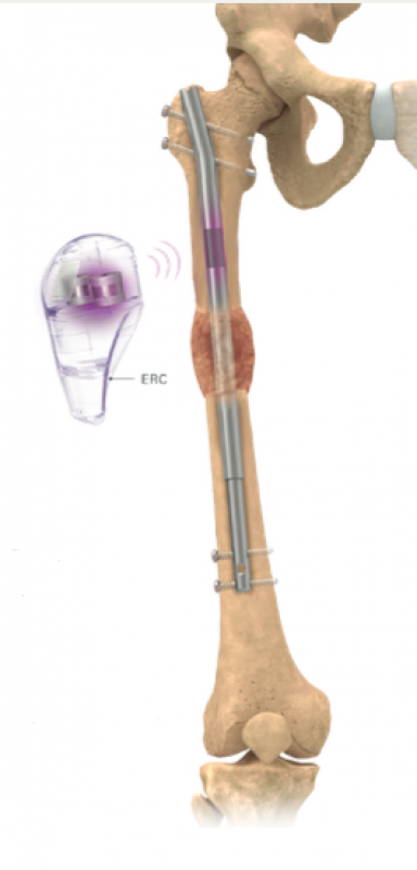
Internal lengthening consists of placing a special lengthening nail inside the bone. The Precice nail (Precice Lengthening System, NuVasive Specialized Orthopedics, San Diego, USA) consists of a metal telescopic rod (so-called lengthening nail) equipped with a mechanism powered by a magnetic field. The nail is placed inside the intramedullary canal through a small skin incision. It is equipped with a small motor supplied by the magnetic field. The motor is activated using an external remote controller (ERC), which allows for the gradual lengthening of the Precice nail. Inside the device, there is a rotating magnet. ERC placed on the designated area on the leg or arm being lengthened and is turned on. The movement of the magnet in the controller results in a slow lengthening of the Precice nail and bone lengthening (it is possible due to locking of the nail ends).

The Precice nail is available in several lengths and diameters, but bones of some people may be still too small to use it. The nail must fit in the intramedullary canal, so the bone diameter must be large enough. The diameter of a specific bone is measured using an X-ray scan. The Precice nail is available in three diameters: 8.5 mm, 10.7 mm and 12.5 mm.
The bone also has to be sufficiently long. The shortest tibial nail is 160 mm long, while the shortest femoral nail is 170 mm long. Based on an X-ray scan, the physician measures the length and verifies if the bone is long enough for receiving a Precice nail.
Another potential problem is the distance between the ERC and the Precice nail. The soft tissues layer (muscle, adipose, skin tissue) can be thick enough to prevent placing the ERC close enough to the Precice nail as to activate the lengthening mechanism. To determine if the Precice system is suitable for an individual patient, the physician measures the distance from the bone’s intramedullary canal to the skin, using the X-ray scan.
It should be noted that patients with a pacemaker and persons planning to undergo magnetic resonance imaging are not eligible for the Precice procedure. Generally, magnetic resonance imaging is not recommended during the treatment using the Precice nail. Therefore, we normally recommend the removal of the apparatus approximately a year after the surgery, provided that the lengthened bone has healed fully.
The maximum length depends on the Precice nail used. Different sizes allow for lengthening the bone by 30, 50 or 80 mm in total. For instance, if only the shortest Precice nail fits into a bone, it can be lengthened by up to 30 mm. In the majority of cases, it is possible to implant a nail allowing for lengthening the bone by 50–80 mm.
The bone is lengthened gradually every day until the planned target length is reached.
The Precice nail can be implanted into the femur of a child from the age of approximately 8. In the case of the tibia, it is recommended to wait until the child reaches the approximate bone maturation age (14 years for girls and 16 years for boys).
PRECICE NAIL IMPLANTATION
The physician has to prepare the place inside the bone where the nail is to be placed. The external bone layer is dense and hard; inside, there is a soft tissue called bone marrow. Using a special drill, the surgeon creates a canal inside the intramedullary cavity in the middle part of the bone, suitable for receiving the chosen Precice nail size.
The nail advancement method depends on the bone to be lengthened and on the individual patient characteristics.
In the case of the femur, the device may be advanced in two ways:
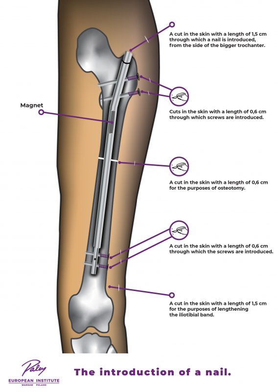
- Distally, i.e. from the pelvis side. Advancing the nail in the direction from the hip towards the knee is called the antegrade (classic) method. The image below shows the nail advancement method, the skin incisions made, as well as the area where the bone is cut (osteotomy) and later lengthened.

Fig. Advancement of the nail into the femur using the antegrade method – the nail is advanced in the direction from the hip through a 2.5 cm incision. Additionally, incisions of approximately 0.5 cm are made, through which the locking screws (two in the proximal and two in the distal part of the nail) are advanced; the same incision is also made for osteotomy. A 2.5 cm incision is made near the knee joint to lengthen the fascia lata.
- Alternatively, proximally, i.e. in the direction from the bottom of the femur, i.e. the knee. Advancing the nail in the direction from the knee towards the hip is called the retrograde method. The image below shows the nail advancement method and the location of the skin incisions and the bone cut (osteotomy). Please note that depending on the advancement method (antegrade or retrograde), the Precice nail magnet is placed in a different area. This affects the place on the thigh where the patient has to place the ERC. When the controller is turned on and placed on the indicated body area, it causes the slow lengthening of the Precice nail.
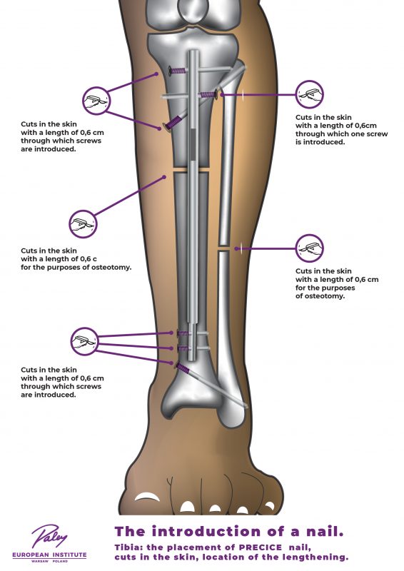
In the case of the tibia, the nail is almost always advanced distally, i.e. in the direction from the knee. The image below shows the nail advancement method, the skin incisions made, as well as the area where the tibia and fibula is cut (osteotomy) and later lengthened. An animation of surgical technique is available.

Fig. Advancement of the nail into the tibia in the direction from the knee through a 2.5 cm incision. Additionally, incisions of approximately 0.5 cm are made, through which the locking screws (three in the proximal and three in the distal part of the nail) are advanced. From the medial side, an incision of approximately 0.5 cm is made for the tibial osteotomy; from the lateral side, an incision of about 2.5 cm is made for the fibular osteotomy.
In the case of the humerus, the nail is advanced from top down, i.e. in the direction from the shoulder towards the elbow.
AFTER THE PROCEDURE
After the Precice nail implementation, the patient stays at the hospital for 2–3 days. Additionally, our specialists recommend the patient not to leave Warsaw for at least the first two weeks following hospital discharge. As the Precice nail is lengthened using the ERC (i.e. during the so-called distraction phase), the patient will return to our site for follow-up visits approximately every 10–14 days. The completed distraction phase is followed by the consolidation phase, during which the bone remodels and heals. During that phase, the patient comes to the site every month until the bone is completely healed.
Physical and occupational therapy plays a very important role during the distraction and consolidation phases. During therapy, the muscles, tendons and ligaments are stretched to ensure full range of motion in the limbs when the treatment ends. Physiotherapy is required in the case of lower limb lengthening and occupational therapy is indicated in the case of humerus lengthening. The physician may allow for the therapy to be completed in the patient’s place of residence, but some cases require specialist rehabilitation provided at our site. Such patients have to stay in Warsaw for the entire therapy duration. In many cases we also use special orthoses/splints. These are usually prepared for the patient in the first week post-surgery.
The patient must not allow full weight-bearing on the leg which has Precice nail implanted. The physician specifies the allowed weight-bearing range for the limb depending on the individual patient’s condition and the size of the nail implanted. If both legs are lengthened, the patient will have to use a wheelchair.
The treating physician must be informed about any planned dentist visits – regardless of whether the visit purpose is routine cleaning or other procedures. After the Precice nail implementation, any dental interventions may require the administration of special antibiotics.
PRECICE NAIL REMOVAL
The Precice nail is usually removed about one year after implementation. The procedure is performed in the outpatient setting, which means that the patient does not have to stay at the hospital overnight. Before deciding to remove the nail, the lengthened bone should be fully healed.
